当教室では以下の内容を中心に研究を進めています。
また平成29年4月より先端画像開発講座(詳細は下記 寄付講座の項をご参照ください)が開設され、協力体制をとりながら、さらに先進的な内容の研究も推進しています。
研究
- 放射線治療における再酸素化とエネルギー代謝の解明
- molecular imagingを用いた放射線治療
- 転移性骨腫瘍に対する集学的治療における放射線治療の最適化の検討
- 体幹部腫瘍に対する3次元放射線治療の有害事象と線量容積因子の検討
- 頭部、頭頸部領域での定位放射線治療の臨床効果および治療計画の最適化
- 前立腺癌に対するI-125永久挿入療法の臨床効果および治療計画の最適化
- 肝・胆・膵の画像診断
- 肝癌の集学的治療
- 血管新生と肝イメージング
- 核医学による悪性腫瘍診断/脳機能測定
- コンピューター支援画像診断(CAD)
- IVR治療に対する画像支援
寄付講座(先端画像開発講座)
スタッフ
特任准教授 兵藤文紀 Fuminori HYODO
特任助教 野澤麻枝 Asae NOZAWA
研究内容
近年の生命科学・分子イメージング技術の進歩により腫瘍内に放射線抵抗性を示す慢性的な低酸素領域や、低酸素と常酸素を繰り返す周期的低酸素領域の存在が明らかとなり、それに伴う放射線治療・診断技術の新たな問題点が浮き彫りになってきました。例えば放射線抵抗性を示す広範な低酸素領域の存在がその一例です。化学療法や放射線治療の治療効果はCTやMRIなどの組織の形態情報に基づく画像診断が適用されていますが、腫瘍サイズに基づく診断には数か月を要するため、効果が得られない場合には患者は大きな時間的損失を被ります。もし化学療法や放射線の治療効果を早期に予測できれば、より効果の高い治療法の選択や副作用の軽減に大いに貢献できると考えられます。
先端画像開発講座では「超偏極=Hyperpolarization」とよばれるMRIの感度を劇的に上昇させる方法を用いて、従来法では可視化が不可能であった生体内分子やレドックス分子を活用した代謝イメージング研究を推進しています。 超偏極技術には様々な方法がありますが、特にフリーラジカルのエネルギ-を利用してMRIの感度を向上する動的確偏極(Dynamic Nuclear Polarization)を応用した生体代謝イメージングを推進していきます。我々は本技術の基礎研究~臨床応用まで展開するトレンスレーショナルリサーチを推進し、病気の早期画像診断や治療効果の早期予測へ役立てたいと考えています。
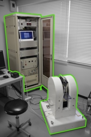
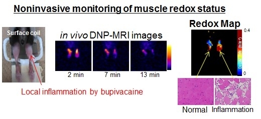
左図 動物用DNP-MRIシステム 右図 炎症(筋炎)のレドックス代謝イメージング
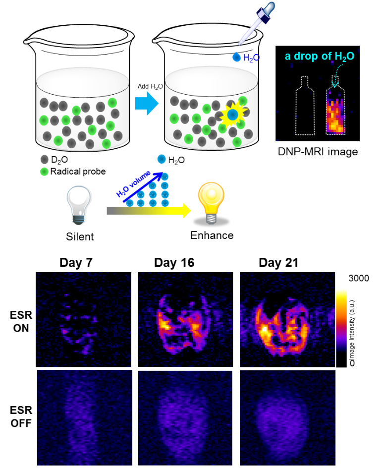
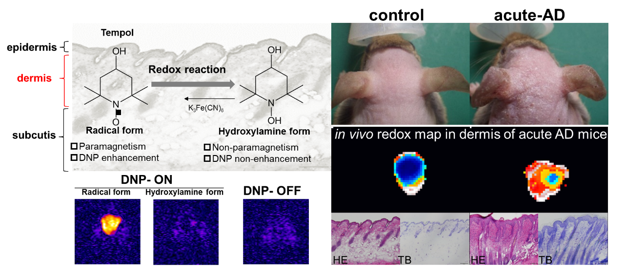
左図 悪性腹水浸潤の超高感度生体イメージング 右図 アトピー性皮膚炎モデルのDNP-MRIによる早期レドックス変動の可視化
欧文論文業績(2018-2020年)※2017年以前は各リンク先をご確認ください。
2022年
2021年
2020年
- Kato H, Kawaguchi M, Ando T, Kaneko Y, Hyodo F, Matsuo M. Hypointense head and neck lesions on T2-weighted images: correlation with histopathologic findings. Neuroradiology. 2020;62(10):1207-1217.
- Makita C, Okada S, Kajiura Y, Tanaka O, Asahi Y, Yamada N, Yanagida M, Kumagai M, Murase S, Ito M, Kumano T, Matsuo M. Vascular events from carotid artery atherosclerosis after radiation therapy for laryngeal and hypopharyngeal cancer: the incidence and risk factors. Nagoya J Med Sci. 2020;82(4):747-761.
- Kawada H, Sato Y, Inaba Y, Yamaura H, Kato M, Murata S, Hasegawa T, Ogura Y, Soga N, Arai Y. Stenting Using the Rendezvous Technique for Postoperative Ureteral Complications in Cancer Patients.Cardiovasc Intervent Radiol. 2020;43(10):1486-1491.
- Noda Y, Goshima S, Okuaki T, Akamine Y, Kajita K, Kawai N, Kawada H, Tanahashi Y, Matsuo M. Hepatocyte fraction: correlation with noninvasive liver functional biomarkers. Abdom Radiol (NY). 2020;45(1):83-89.
- Noda Y, Goshima S, Nagata S, Kawada H, Tanahashi Y, Kato T, Suwa T, Kawai N, Yabe D, Matsuo M. Utility of microcatheter in adrenal venous sampling for primary aldosteronism. Br J Radiol. 2020;93(1109):20190636.
- Noda Y, Goshima S, Tsuji Y, Kajita K, Akamine Y, Kawai N, Kawada H, Tanahashi Y, Matsuo M. Pancreatic extracellular volume fraction using T1 mapping in patients with impaired glucose intolerance. Abdom Radiol (NY). 2020;45(2):449-456.
- Noda Y, Goshima S, Takai Y, Kawai N, Kawada H, Tanahashi Y, Matsuo M. Detection of pancreatic ductal adenocarcinoma and liver metastases: comparison of Gd-EOB-DTPA-enhanced MR imaging vs. extracellular contrast materials. Abdom Radiol (NY). 2020;45(8):2459-2468.
- Noda Y, Goshima S, Nakashima Y, Miyoshi T, Kawai N, Kambadakone A, Matsuo M. Iodine dose optimization in portal venous phase virtual monochromatic images of the abdomen: Prospective study on rapid kVp switching dual energy CT. Eur J Radiol. 2020;122:108746.
- Noda Y, Goshima S, Kaga T, Ando T, Miyoshi T, Kawai N, Kawada H, Tanahashi Y, Matsuo M. Virtual monochromatic image at lower energy level for assessing pancreatic ductal adenocarcinoma in fast kV-switching dual-energy CT. Clin Radiol. 2020;75(4):320.e17-320.e23.
- Ando T, Kato H, Kawaguchi M, Tanahashi Y, Aoki M, Kuze B, Matsuo M. Diagnostic ability of contrast-enhanced computed tomography for metastatic cervical nodes in head and neck squamous cell carcinomas: significance of additional coronal reconstruction images. Pol J Radiol. 2020;85:e1-e7.
- Ando T, Kato H, Kawaguchi M, Furui T, Morishige KI, Hyodo F, Matsuo M. MR findings for differentiating decidualized endometriomas from seromucinous borderline tumors of the ovary. Abdom Radiol (NY). 2020;45(6):1783-1789.
- Kawaguchi M, Kato H, Tomita H, Hara A, Matsuo M. CT and MR imaging findings of solitary nevus lipomatosus cutaneous superficialis: radiological-pathological correlation. Skeletal Radiol. 2020;49(1):129-135.
- Kawaguchi M, Kato H, Tomita H, Hara A, Suzui N, Miyazaki T, Matsuyama K, Seishima M, Matsuo M. Magnetic Resonance Imaging Findings Differentiating Cutaneous Basal Cell Carcinoma from Squamous Cell Carcinoma in the Head and Neck Region. Korean J Radiol. 2020;21(3):325-331.
- Kawaguchi M, Kato H, Hatano Y, Tomita H, Hara A, Suzui N, Miyazaki T, Furui T, Morishige K, Matsuo M. MR imaging findings of low-grade serous carcinoma of the ovary: comparison with serous borderline tumor. Jpn J Radiol. 2020;38(8):782-789.
- Kawaguchi M, Kato H, Tomita H, Hara A, Suzui N, Miyazaki T, Matsuo M. Comparison of Imaging Findings between Human Papillomavirus-positive and -Negative Squamous Cell Carcinomas of the Maxillary Sinus. J Clin Imaging Sci. 2020;10:59.
- Kawaguchi M, Kato H, Kaneko Y, Matsuo M. Hyperdense Thymic Atrophy After Chemotherapy in Pediatric Patients With Extrathoracic Malignancies. J Comput Assist Tomogr. 2020;44(6):865-869.
- Kawaguchi M, Kato H, Tomita H, Hara A, Suzui N, Miyazaki T, Matsuyama K, Seishima M, Matsuo M. MR imaging findings for differentiating cutaneous malignant melanoma from squamous cell carcinoma. Eur J Radiol. 2020;132:109212.
- Kaga T, Kato H, Hatano Y, Kawaguchi M, Furui T, Morishige KI, Matsuo M. Can MRI features differentiate ovarian mucinous carcinoma from mucinous borderline tumor? Eur J Radiol. 2020;132:109281.
- Tanaka O, Sugiyama A, Omatsu T, Tawada M, Makita C, Matsuo M. Hemostatic radiotherapy for inoperable gastric cancer: a pilot study. Br J Radiol. 2020;93(1111):20190958.
- Tanaka O, Kojima T, Ohbora A, Makita C, Taniguchi T, Ono K, Matsuo M, Nagata Y. Scores of Child-Pugh Classification Impact Overall Survival After Stereotactic Body Radiation Therapy for Primary and Metastatic Liver Tumors. J Clin Exp Hepatol. 2020;10(2):101-105.
- Tanaka O, Ono K, Taniguchi T, Makita C, Matsuo M. Dosimetric evaluation of the heart and left anterior descending artery dose in radiotherapy for Japanese patients with breast cancer. J Radiat Res. 2020;61(1):134-139.
- Tanaka O, Funaguchi N, Toyoshi S, Taniguchi T, Ono K, Kunishima Y, Matsuo M. Biologically effective dose and overall survival in stereotactic body radiotherapy for lung tumors. Magazine of Medical Oncology. 2020;13:353-356.
- Tanaka O, Taniguchi T, Kuroki K, Ono K, Makita C, Matsuo M. Pickup effect of patients with potential radiotherapy indication by Radiographers: an institutional reports in Japan. Oncology and Radiotherapy. 2020;52:089-091.
- Tanaka O, Ono K, Taniguchi T, Omatsu T, Sakamoto N, Makita C, Matsuo M. Hemostasis irradiation for gastric cancer: Comparison of whole and partial stomach irradiation in the liver and kidney. Radiologica I Therapia. 2020;14(6).
- Tanaka H, Makita C, Manabe Y, Kajima M, Matsuyama K, Matsuo M. Radiation therapy combined with bone-modifying agents ameliorates local control of osteolytic bone metastases in breast cancer. J Radiat Res. 2020;61(3):494-498.
- Tanahashi Y, Iwasaki R, Shoda S, Kawada H, Ando T, Takasu M, Hyodo F, Goshima S, Mori T, Matsuo M. Dynamic contrast-enhanced computed tomography lymphangiography with intranodal injection of water-soluble iodine contrast media in microminipig: imaging protocol and feasibility. Eur Radiol. 2020;30(11):5913-5922.
- Tanahashi Y, Kawada H, Goshima S, Takahashi T, Yoshida K, Matsuo M. Intranodal Popliteal Lymphangiography for Postoperative Lymphorrhea after Inguinal Node Dissection. J Vasc Interv Radiol. 2020;31(11):1926-1929.
- Tanahashi Y, Ozeki M, Kawada H, Goshima S, Fukao T, Matsuo M. Direct-Puncture Lymphatic Embolization in the Prone Position for Chylothorax Caused by Lymphatic Anomaly. J Vasc Interv Radiol. 2020;31(5):849-852.e1.
- Shoda S, Hyodo F, Tachibana Y, Kiniwa M, Naganuma T, Eto H, Koyasu N, Murata M, Matsuo M. Imaging of Hydroxyl-Radical Generation Using Dynamic Nuclear Polarization-Magnetic Resonance Imaging and a Spin-Trapping Agent. Anal Chem. 2020;92(21):14408-14414.
- Taniguchi T, Iinuma K, Kato D, Takai M, Maekawa YM, Nakane K, Mizutani K, Tsuchiya T, Nakano M, Kato T, Ito M, Kumano T, Matsuo M, Koie T. Predictive factors of rectal hemorrhage in patients with localized prostate cancer who underwent low-dose-rate brachytherapy. Int J Clin Oncol. 2020;25(9):1711-1717.
- Miyai M, Kanayama T, Hyodo F, Kinoshita T, Ishihara T, Okada H, Suzuki H, Takashima S, Wu Z, Hatano Y, Egashira Y, Enomoto Y, Nakayama N, Soeda A, Yano H, Hirata A, Niwa M, Sugie S, Mori T, Maekawa Y, Iwama T, Matsuo M, Hara A, Tomita H. Glucose transporter Glut1 controls diffuse invasion phenotype with perineuronal satellitosis in diffuse glioma microenvironment. Neurooncol Adv. 2020;3(1):vdaa150.
- Urano M, Nishikawa H, Goto T, Shiraki N, Matsuo M, Denewar FA, Kondo N, Toyama T, Shibamoto Y. Digital Mammographic Features of Breast Cancer Recurrences and Benign Lesions Mimicking Malignancy Following Breast-Conserving Surgery and Radiation Therapy. Kurume Med J. 2020;65(4):113-121.
- Murai T, Matsuo M, Tanaka H, Manabe Y, Takaoka T, Hachiya K, Yamaguchi T, Otsuka S, Shibamoto Y. Efficacy of herbal medicine TJ-14 for acute radiation-induced enteritis: a multi-institutional prospective Phase II trial. J Radiat Res. 2020;61(1):140-145.
- Iinuma K, Nakano M, Kato T, Kato D, Takai M, Maekawa YM, Nakane K, Mizutani K, Tsuchiya T, Ishihara T, Ito M, Matsuo M, Koie T. Assessment of Long-term Changes in Lower Urinary Tract Symptoms in Patients With Prostate Cancer Who Underwent Low-dose-rate Prostate Brachytherapy. Urology. 2020;142:213-220.
- Ninomiya H, Ozeki M, Matsuzawa Y, Nozawa A, Yasue S, Kubota K, Endo S, Asano T, Taguchi K, Ohe N, Matsuo M, Iwama T, Ohnishi H. A pediatric case of anaplastic astrocytoma with a gliomatosis cerebri; the growth pattern and changes in serum VEGF-121 levels after bevacizumab treatment. J Clin Neurosci. 2020;81:431-433.
- Yasuda T, Tanaka O, Hayashi S, Nakahata Y, Yasuda Y, Omatsu T, Obora A, Kojima T, Matsuo M, Yagi N. Successful treatment of unresectable advanced rectal cancer with liver metastases by hemostasis re-irradiation of the rectal cancer and palliative low-dose whole-liver radiation therapy: a case report. Clin J Gastroenterol. 2020;13(1):11-16.
- Ohashi T, Terasawa K, Aoki M, Akazawa T, Shibata H, Kuze B, Asano T, Kato H, Miyazaki T, Matsuo M, Inoue N, Ito Y. The importance of FDG-PET/CT parameters for the assessment of the immune status in advanced HNSCC. Auris Nasus Larynx. 2020;47(4):658-667.
- Ishida K, Tomita H, Kanayama T, Noguchi K, Niwa A, Kawaguchi M, Miyai M, Matsuo M, Imaizumi Y, Kato K, Hatano Y, Hirata A, Okada H, Shibata T, Hara A. Specific Deletion of p16INK4a with Retention of p19ARF Enhances the Development of Invasive Oral Squamous Cell Carcinoma. Am J Pathol. 2020;190(6):1332-1342.
- Numasaki H, Teshima T, Ando Y, Akuta K, Ikeda H, Okajima K, Kumano T, Sasaki T, Sekiguchi K, Tago M, Terahara A, Nakamura K, Nishimura T, Ogawa K; for Society Japanese Radiation Oncology Database Committee Japanese structure survey of radiation oncology in 2012. J Radiat Res. 2020;61(1):146-160.
- Kagiya G, Sato A, Ogawa R, Hatashita M, Kato M, Kubo M, Kojima F, Kawakami F, Nishimura Y, Abe N, Hyodo F. Real-time visualization of intratumoral necrosis using split-luciferase reconstitution by protein trans-splicing.Mol Ther Oncolytics. 2020;20:48-58.
- Hosain MZ, Hyodo F, Mori T, Takahashi K, Nagao Y, Eto H, Murata M, Akahoshi T, Matsuo M, Katayama Y. Development of a novel molecular probe for the detection of liver mitochondrial redox metabolism. Sci Rep. 2020;10(1):16489.
- Nakamura K, Ishikawa H, Akimoto T, Aoki M, Kariya S, Kawamura H, Kumano T, Kozuka T, Konishi K, Sakaguchi M, Takayama K, Other authors are shown in Appendix National survey of radiation oncologists' practice patterns regarding hormone-naïve prostate cancer with bone metastases. Jpn J Clin Oncol. 2020;50(10):1188-1194.
- Sakurai T, Takamatsu S, Shibata S, Iwata K, Taka M, Gabata T, Kumano T, Makino T, Mizokami A. Toxicity and clinical outcomes of single-fraction high-dose-rate brachytherapy combined with external beam radiotherapy for high-/very high-risk prostate cancer: A dosimetric analysis of toxicity. Jpn J Radiol. 2020;38(12):1197-1208.
- Ide M, Sonoda N, Inoue T, Kimura S, Minami Y, Makimura H, Hayashida E, Hyodo F, Yamato M, Takayanagi R, Inoguchi T. The dipeptidyl peptidase-4 inhibitor, linagliptin, improves cognitive impairment in streptozotocin-induced diabetic mice by inhibiting oxidative stress and microglial activation. PLoS One. 2020;15(2):e0228750.
- Mu Y, Li J, Kang JH, Eto H, Zai K, Kishimura A, Hyodo F, Mori T, Katayama Y. A Lipid-Based Nanocarrier Containing Active Vitamin D3 Ameliorates NASH in Mice via Direct and Intestine-Mediated Effects on Liver Inflammation. Biol Pharm Bull. 2020;43(9):1413-1420.
- Suetsugu T, Tanaka Y, Banno S, Fukada M, Yasufuku I, Iwata Y, Imai T, Tanahashi T, Matsui S, Imai H, Matsuhashi N, Takahashi T, Yamaguchi K, Tanahashi Y, Kawada H, Matsuo M, Yoshida K. Intranodal lymphangiography for chyle leakage after esophagectomy: A case report. Mol Clin Oncol. 2020;12(4):343-349.
- Kamiya N, Oshima A, Zhou X, Kato H, Hara T, Miyoshi T, Matsuo M, Fujita H. Surface Muscle Segmentation Using 3D U-Net Based on Selective Voxel Patch Generation in Whole-Body CT Images. Applied Sciences. 2020;10:4477.
- Kato H, Kawaguchi M, Ando T, Kaneko Y, Hyodo F, Matsuo M. Hypointense head and neck lesions on T2-weighted images: correlation with histopathologic findings. Neuroradiology. 2020;62(10):1207-1217.
2019年
- Kato H, Esaki K, Yamaguchi T, Tanaka H, Kajita K, Furui T, Morishige KI, Goshima S, Matsuo M. Predicting Early Response to Chemoradiotherapy for Uterine Cervical Cancer Using Intravoxel Incoherent Motion MR Imaging. Magn Reson Med Sci. 2019; 18(4):293-298.
- Kaneko Y, Kato H, Matsuo M. Hilar and mediastinal sarcoid-like reaction after the treatment of malignant tumors: imaging features and natural course on 18F-FDG-PET/CT.Jpn J Radiol. 2019; 37(1):88-94.
- Kawaguchi M, Kato H, Aoki M, Kuze B, Hara A, Matsuo M. CT and MR imaging findings of infection-free and benign second branchial cleft cysts. Radiol Med. 2019; 124(3):199-205.
- Kawaguchi M, Kato H, Suzui N, Furui T, Morishige KI, Goshima S, Matsuo M. MR imaging findings differentiating uterine submucosal polypoid adenomyomas from endometrial polyps. Br J Radiol. 2019; 92(1095):20180430.
- Nozawa A, Ozeki M, Hori T, Kato H, Ohe N, Fukao T. Fatal progression of Gorham-Stout disease with skull base osteomyelitis and lateral medullary syndrome. Intern Med. 2019; 58:1929-1933.
- Ishida K, Kato K, Inoue K, Hatakeyama D, Kato H, Shibata T. A case of herniation of the mylohyoid muscle with penetration of the sublingual gland. J Oral Maxillofac Surg Med Pathol.2019; 31(3):189-191.
- Kawaguchi M, Kato H, Nakano M, Goshima S, Matsuo M. Clinical features of bone metastasis with extraosseous soft-tissue mass in prostate cancer patients. Br J Radiol open. 2019; 1:20180042.
- Nishibori H, Kato H, Kawaguchi M, Nagano A, Matsuo M. T2*-weighted MR imaging findings of giant cell tumors of bone: radiological-pathological correlation. Jpn J Radiol. 2019; 37(6):473-480.
- Fujimoto K, Kato H, Kaneko Y, Aoki M, Kuze B, Kato K, Shibata T, Matsuo M. Clavicle fracture following neck dissection: imaging features and natural course. Br J Radiol. 2019; 92(1100):20190054.
- Kawaguchi M, Kato H, Matsuo M. CT and MRI features of scalp lesions. Radiol Med. 2019; 124(10):1049-1061.
- Kawaguchi M, Kato H, Goshima S, Matsuo M. Response to Pilomatricoma (calcifying epithelioma): MDCT and MR imaging findings in 31 patients with radiological-pathological correlation. Eur J Radiol. 2019; 118:293.
- Kawaguchi M, Kato H, Hara A, Suzui N, Tomita H, Miyazaki T, Iwata H, Matsuo M. CT and MRI characteristics for differentiating mediastinal Müllerian cysts from bronchogenic cysts. Clin Radiol. 2019; 74(12):976.e19-976.e25.
- Iwashita T, Uemura S, Shimizu M, Hyodo F, Tomita H, Iwasaki R, Takasu M, Mori T, Tanaka H,
- Matsuo M. Endoscopic Ultrasound-Guided Fine-Needle Injection of Hydrogen Peroxide into the Pancreas: Feasibility and Tolerability Study Using a Survival Porcine Model. Ultrasound Med Biol. 2019 Feb; 45(2):579-585
- Hyodo F, Naganuma T, Eto H, Murata M, Utsumi H, Matsuo M. In vivo melanoma imaging based on dynamic nuclear polarization enhancement in melanin pigment of living mice using in vivo dynamic nuclear polarization magnetic resonance imaging. Free Radic Biol Med. 2019; 134:99-105.
- Cui Y, Masaki K, Zhang X, Yamasaki R, Fujii T, Ogata H, Hayashida S, Yamaguchi H, Hyodo F,
- Eto H, Koyama S, Iinuma K, Yonekawa T, Matsushita T, Yoshida M, Yamada K, Kawano M, Malissen M, Malissen B, Kira J. A novel model for treatment of hypertrophic pachymeningitis. Ann Clin Transl Neurol. 2019; 6(3):431-444.
- Kodama Y, Hyodo F, Yamato M, Yasukawa K, Minami Y, Sonoda N, Ogawa Y, Ichikawa K, Inoguchi T. Dynamic nuclear polarization magnetic resonance imaging and the oxygen-sensitive paramagnetic agent OX63 provide a noninvasive quantitative evaluation of kidney hypoxia in diabetic mice. Kidney Int. 2019; 96(3):787-792.
- Mitsuzaki K, Iinuma G, Morimoto T, Miyake M, Tomimatsu H.Computed tomographic colonography with a reduced dose of laxative using a novel barium sulfate contrast agent in Japan. Japanese journal of radiology. 2019; 37(3):245-254.
- Noda Y, Goshima S, Tsuji Y, Tomita H, Hara A, Kawaguchi M, Kawada H, Kawai N, Tanahashi Y,
- Matsuo M. Prognostic evaluation of pancreatic ductal adenocarcinoma: Associations between molecular biomarkers and CT imaging findings. Pancreatology. 2019 Mar; 19(2):331-339.
- Noda Y, Goshima S, Suzui N, Miyazaki T, Kajita K, Kawada H, Kawai N, Tanahashi Y, Matsuo M. Pancreatic MRI associated with pancreatic fibrosis and postoperative fistula: comparison between pancreatic cancer and non-pancreatic cancer tissue. Clin Radiol. 2019 Jun; 74(6):490.e1-490.e6.
- Noda Y, Goshima S, Tsuji Y, Kajita K, Kawada H, Kawai N, Tanahashi Y, Matsuo M. Correlation of quantitative pancreatic T 1 value and HbA1c value in subjects with normal and impaired glucose tolerance. J Magn Reson Imaging. 2019; 49(3):711-718.
- Kawai N, Goshima S, Noda Y, Kajita K, Kawada H, Tanahashi Y, Nagata S, Matsuo M. Gadoxetic acid-enhanced dynamic magnetic resonance imaging using optimized integrated combination of compressed sensing and parallel imaging technique. Magn Reson Imaging. 2019; 57:111-117.
- Nagata S, Goshima S, Noda Y, Kawai N, Kajita K, Kawada H, Tanahashi Y, Matsuo M. Magnetic resonance cholangiopancreatography using optimized integrated combination with parallel imaging and compressed sensing technique. Abdominal Radiology. 2019; 44(5):1766-1772.
- Tanaka H, Nakashima Y, Ito M, Yamaguchi T, Esaki K, Kamei S, Ishihara S, Hayashi M, Ogawa S, Goshima S, Matsuo M. Intensity-modulated radiation therapy for elderly patients (aged ≥75 years) with localized prostate cancer: Comparison with younger patients (aged <75 years). Mol Clin Oncol. 2019; 10(4):476-480.
- Tanaka H, Kawaguchi M, Shoda S, Miyoshi T, Iwasaki R, Hyodo F, Mori T, Hara A, Tomita H, Matsuo M. Nuclear Accumulation of β-Catenin in Cancer Stem Cell Radioresistance and Stemness in Human Colon Cancer. Anticancer Res. 2019 Dec; 39(12):6575-6583.
- Koyasu H, Goshima S, Noda Y, Nishibori H, Takeuchi M, Matsunaga K, Yamada T, Matsuo M. The feasibility of dedicated breast PET for the assessment of residual tumor after neoadjuvant chemotherapy. Jpn J Radiol. 2019; 37(1):81-87.
- Tanaka O, Yokoi R, Mukai T, Yamada M, Kato T, Taniguchi T, Ono K, Matsuo M. Radiotherapy for gastric bleeding from tumor invasion of recurrent colon cancer with liver metastasis after resection. J Gastrointest Cancer. 2019; 50(2): 349-352.
- Tanaka O, Omatsu T, Kariya S, Maejima R, Taniguchi T, Ono K, Kunishima Y, Matsuo M. Usefulness of diffusion-weighted magnetic resonance imaging for evaluating the effect of hemostatic radiotherapy for unresectable gastric cancer. Clin J Gastroenterol. 2019; 12(3):269-273.
- Tanaka O, Kunishima Y, Taniguchi T, Ono K, Matsuo M. Changes in patients' quality of life during radiotherapy and 1 month after treatment. Magazine of European Medical Oncology. 2019; 12:77-82.
- Tanaka O, Seike K, Taniguchi T, Ono K, Matsuo M. Investigation of the changes in the prostate, bladder, and rectal wall sizes during external beam radiotherapy. Reports of Practical Oncology and Radiotherapy. 2019; 24(2):204-207.
- Tanaka O, Masui Y, Matsunami R, Kawase S, Ono K, Taniguchi T, Kawaguchi Y, Matsuo M. Comparison of Conventional 2D CC (Cranio-Caudal) + MLO (Medio-Lateral Oblique) Bi-Directional Photography and 2D-MLO + DBT-MLO (Digital Breast Tomosynthesis) in Mammography Examination. Archives of Radiology. 2019; 2:11.
- Tanaka O, Matsuura K, Sugiyama A, Kato T, Tomita E, Matsuo M. Hemostatic Radiotherapy Used Twice for Inoperable Progressive Gastric Cancer with Bleeding. J Gastrointest Cancer. 2019; 50(1):151-155.
- Tanaka O, Miki K, Taniguchi T, Kojima T, Ohbora A, Makita C, Matsuo M. 99m-Technetium Galactosyl Human Serum Albumin Scanning to Evaluate Liver Function After Stereotactic Body Radiotherapy for Hepatocellular Carcinoma: A Case Report. Radiology Case Reports. 2019:14(11); 1410-1414
- Kanno M, Narita N, Fujimoto Y, Wakisaka N, Yoshizaki T, Kodaira T, Makita C, Sato Y, Yamazaki K, Wakaoka T, Shimode Y, Tsuji H, Kito R, Ishinaga H, Hosokawa S, Takakura H, Nishimura K, Matoba T, Fujieda S. Third Epidemiological Analysis of Nasopharyngeal Carcinoma in the Central Region of Japan from 2006 to 2015. Cancers (Basel). 2019; 11(8):1180.
- Shimizu H, Sasaki K, Ito M, Aoyama T, Tachibana H, Tomita N, Makita C, Tanaka H, Koide Y, Iwata T, Kodaira T. Impact of treatment planning using a structure block function on the target and organ doses related to patient movement in cervical esophageal cancer: A phantom study. J Appl Clin Med Phys. 2019; 20(5):75-83.
- Iinuma K, Mizutani K, Kato T, Nakane K, Tanaka H, Nakano M, Matsuo M, Koie T. Spontaneous healing of rectal penetration by SpaceOAR ® hydrogel insertion during permanent iodine-125 implant brachytherapy: A case report. Mol Clin Oncol. 2019; 11(6):580-582.
- Hara T, Niwa S, Urikura A, Matsubara K, Hoshino T, Nishimaru E, Taniguchi T. Assessment of longitudinal beam property and contrast uniformity for 256‐ and 320‐row area detector computed tomography scanners in the 160‐mm nonhelical volume‐acquisition mode. JACMP. 2019; 20(8): 164-170.
- Kajita K, Goshima S, Noda Y, Kawada H, Kawai N, Okuaki T, Honda M, Matsuo M. Thin-slice Free-breathing Pseudo-golden-angle Radial Stack-of-stars with Gating and Tracking T 1-weighted Acquisition: An Efficient Gadoxetic Acid-enhanced Hepatobiliary-phase Imaging Alternative for Patients with Unstable Breath Holding. Magn Reson Med Sci. 2019 10;18(1):4-11.
- Kato H, Kawaguchi M, Ando T, Aoki M, Kuze B, Matsuo M. CT and MR Imaging Findings of Non-Neoplastic Cystic Lesions of the Parotid Gland. Jpn J Radiol. 2019; 37(9):627-635.
2018年
- Matsuo M, Kawai T, Kishimoto S, Saito K, Munasinghe J, Devasahayam N, Mitchell JB, Krishna MC. Co-imaging of the tumor oxygenation and metabolism using electron paramagnetic resonance imaging and 13-C hyperpolarized magnetic resonance imaging before and after irradiation. Oncotarget. 2018;9(38):25089-25100.
- Kimura M, Kato H, Sekino S, Ishida N, Murase K, Shimabukuro K, Sekino T, Doi K, Matsuo M. Radical resection of a giant retroperitoneal calcifying fibrous tumor combined with right hepatectomy and reconstruction of the inferior vena cava and bilateral renal veins. Surg Case Rep. 2018;4(1):7.
- Ando T, Kato H, Mochizuki K, Ozawa K, Goshima S, Matsuo M. MR findings of the orbit in patients with Vogt-Koyanagi-Harada disease. Neuroradiology. 2018;60(4):421-426.
- Ando T, Kato H, Furui T, Morishige KI, Goshima S, Matsuo M. Uterine smooth muscle tumours with hyperintense area on T1 weighted images: differentiation between leiomyosarcomas and leiomyomas. Br J Radiol. 2018;91(1084):20170767.
- Kato H, Ozeki M, Fukao T, Matsuo M. Chest imaging in generalized lymphatic anomaly and kaposiform lymphangiomatosis. Pediatr Int. 2018;60(7):667-668.
- Kawaguchi M, Kato H, Miyazaki T, Kato K, Hatakeyama D, Mizuta K, Aoki M, Matsuo M. CT and MR imaging characteristics of histological subtypes of head and neck ossifying fibroma. Dentomaxillofac Radiol. 2018;47(6):20180085.
- Kawaguchi M, Kato H, Hatano Y, Mizuno N, Furui T, Morishige KI, Hara A, Goshima S, Matsuo M. Inchworm sign of endometrial cancer on diffusion-weighted MRI: radiology-pathology correlation. Clin Radiol. 2018;73(10):907.e9-907.e14.
- Enomoto Y, Yamauchi K, Asano T, Otani K, Iwama T. Effect of metal artifact reduction software on image quality of C-arm cone-beam computed tomography during intracranial aneurysm treatment. Interv Neuroradiol. 2018;24(3):303-308.
- Tanahashi Y, Kondo H, Yamamoto M, Osawa M, Yokoyama T, Sugawara T, Kawada H, Goshima S, Matsuo M, Furui S, Oba H. Efficacy of Automated Supplying Artery Tracking Software Using Multidetector-Row Computed Tomography Images for Emergent Transcatheter Arterial Embolization. Cardiovasc Intervent Radiol. 2018;41(11): 1786-1793.
- Tanaka H, Yamaguchi T, Hachiya K, Miwa K, Shinoda J, Hayashi M, Ogawa S, Nishibori H, Goshima S, Matsuo M. 11C-methionine positron emission tomography for target delineation of recurrent glioblastoma in re-irradiation planning. Rep Pract Oncol Radiother. 2018;23:215-219.
- Tanaka H, Yamaguchi T, Hachiya K, Kamei S, Ishihara S, Hayashi M, Ogawa S, Nishibori H, Goshima S, Matuo M. Treatment outcomes and late toxicities of intensity-modulated radiation therapy for 1091 Japanese patients with localized prostate cancer. Rep Pract Oncol Radiother. 2018;23:28-33.
- Tanaka H, Kato A, Kawaguchi M, Yamaguchi T, Kitahara M, Matsuyama K, Okada S, Matsuo M. Pelvic insufficiency fractures after whole pelvic irradiation for uterine cervical cancer. Eur J Gynaecol Oncol. 2018;39:361-364.
- Kawano T, Murata M, Kang JH, Piao JS, Narahara S, Hyodo F, Hamano N, Guo J, Oguri S, Ohuchida K, Hashizume M. Ultrasensitive MRI detection of spontaneous pancreatic tumors with nanocage-based targeted contrast agent. Biomaterials. 2018;152:37-46.
- Matsumoto KI, Hyodo F, Mitchell JB, Krishna MC. Effect of body temperature on the pharmacokinetics of a triarylmethyl-type paramagnetic contrast agent used in EPR oximetry. Magn Reson Med. 2018;79(2):1212-1218.
- Irie M, Hayakawa E, Fujimura Y, Honda Y, Setoyama D, Wariishi H, Hyodo F, Miura D. Analysis of spatiotemporal metabolomic dynamics for sensitively monitoring biological alterations in cisplatin-induced acute kidney injury. Biochem Biophys Res Commun. 2018;496(1):140-146.
- Hyodo F, Sho T, Maity B, Fujita K, Tachibana Y, Akashi S, Mano M, Hishikawa Y, Matsuo M, Ueno T. Photoinduced in Vivo Magnetic Resonance Imaging (MRI) with Rapid CO Release from an MnCO-Protein Needle Composite. Chemistry. 2018;24(45):11578-11583.
- Noda Y, Goshima S, Kozaka K, Yoneda N, Mizuno N, Kato A, Fujimoto K, Tsuji Y, Miyoshi T, Kawada H, Kawai N, Tanahashi Y, Matsuo M. Optimal window settings in single-source dual-energy computed tomography of the abdomen. Eur J Radiol. 2018;109:204-209.
- Noda Y, Goshima S, Miyoshi T, Kawada H, Kawai N, Tanahashi Y, Matsuo M. Assessing Chemotherapeutic Response in Pancreatic Ductal Adenocarcinoma: Histogram Analysis of Iodine Concentration and CT Number in Single-Source Dual-Energy CT. AJR Am J Roentgenol. 2018;211(6):1221-1226.
- Noda Y, Goshima S, Fujimoto K, Kawada H, Kawai N, Tanahashi Y, Matsuo M. Utility of the portal venous phase for diagnosing pancreatic necrosis in acute pancreatitis using the CT severity index. Abdom Radiol (NY). 2018;43(11):3035-3042.
- Noda Y, Goshima S, Kawada H, Kawai N, Miyoshi T, Matsuo M, Bae KT. Modified National Comprehensive Cancer Network Criteria for Assessing Resectability of Pancreatic Ductal Adenocarcinoma. AJR Am J Roentgenol. 2018;210(6):1252-1258.
- Noda Y, Goshima S, Nagata S, Miyoshi T, Kawada H, Kawai N, Tanahashi Y, Matsuo M. Right adrenal vein: comparison between adaptive statistical iterative reconstruction and model-based iterative reconstruction. Clin Radiol. 2018;73(6):594. e1-594. e6.
- Noda Y, Goshima S, Namimoto T, Shinkawa N, Nakagawa M, Kajita K, Kawada H, Kawai N, Tanahashi Y, Matsuo M, Bae KT, Hirai T, Yamashita Y. Simultaneous acquisition of MR angiography and diagnostic images of abdomen at view-sharing multiarterial phases and comparing the effect of two different contrast agents. J Magn Reson Imaging. 2018;48(1):102-110.
- Tanaka O, Komeda H, Tamaki M, Seike K, Fujimoto S, Yama E, Hirose S, Matsuo M. Comparison of MRI visualization between linearly placed iron-containing and non-iron-containing fiducial markers for prostate radiotherapy. Br J Radiol. 2018;91:2017061.Tanaka O, Komeda H, Hirose S, Taniguchi T, Ono K, Yama E, Matsuo M. Influence of gold marker for magnetic resonance imaging during prostate radiotherapy. Polish Journal of Medical Physics and Engineering. 2018;24:99-101.
- Tanaka O, Komeda H, Hirose S, Taniguchi T, Ono K, Matsuo M. Visibility of an iron-containing fiducial marker in magnetic resonance imaging for high-precision external beam prostate radiotherapy. Asia Pac J Clin Oncol. 2018;14:e405-e411.
2017年
2016年
2015年
競争的研究資金獲得状況(2015-2020年度)
過去の競争的研究資金獲得実績(※対象期間の終了したものはこちらをご覧ください。)
研究代表者:兵藤文紀
科学研究費助成事業 基盤研究(B)
「ミトコンドリア代謝をイメージングバイオマーカーとする早期画像診断法の開発」
研究機関:2019年4月1日-2023年3月31日
研究代表者:野澤麻枝
科学研究費助成事業 基盤研究(C)
「乳癌の酸化還元代謝に基づく早期診断法の開発」
研究機関:2019年4月1日-2022年3月31日
研究代表者:松尾政之
科学研究費助成事業 国際共同研究加速基金(国際共同研究強化(B))
「臨床応用を目的とする酸素・代謝を指標とする新たな分子画像診断技術の国際共同開発」
研究期間:2020年 4月 1日-2024年 3月31日
研究代表者:水野 希
科学研究費助成事業 若手研究
「高感度水動態の可視化技術の開発と腹膜播種モデルへの応用」
研究期間:2020年 4月 1日-2022年 3月31日
研究代表者:川田紘資
科学研究費助成事業 若手研究
「医療費削減・安全性向上を実現するRadioPackEmbolizationの開発」
研究期間:2020年 4月 1日-2023年 3月31日
研究代表者:牧田智誉子
日本女性放射線腫瘍医の会・助成事業
「Vascular events from carotid artery atherosclerosis after radiation therapy for laryngeal
and hypopharyngeal cancer: the incidence and risk factors」
日本放射線腫瘍学会 他領域関連学会発表補助事業
臨床研究に関する情報公開
岐阜大学放射線科では、教育研究機関の1部署として、患者さんの診療情報等を研究目的に利用させていただくことがあります(「臨床研究」といいます)。
臨床研究は国が定める「人を対象とする医学系研究に関する倫理指針」に基づいて行っています。臨床研究への協力をお願いする際には、研究の実施方法・内容に応じて、文書若しくは口頭による同意を得るか、予め研究に関する情報をホームページ等で通知・公開し、研究参加可否の機会を提供することになっています。ここでは、後者の方法で実施する臨床研究について、情報を公開しています。
平成30年7月以前の研究課題一覧
| 承認番号 | 研究課題名 | 研究期限 |
|---|---|---|
| 27-500 | 画像下治療(IVR治療)に関する後方視的研究 | 平成33年(令和3年)2月 |
| 29-144 | 全国放射線治療症例に基づく放射線治療の実態調査および質評価 | 平成32年(令和2年)3月 |
| 29-304 | 間質性肺炎合併肺癌の術後間質性肺炎急性憎悪におけるイメージングバイオマーカーの確立 | 平成31年(令和元年)12月 |
| 29-366 | 全国国立大学附属病院におけるCT撮影線量調査 | 平成35年(令和5年)3月 |
| 29-458 | 腹部ステントグラフト内挿術後のタイプⅡエンドリークに対するIVR:技術的側面と予後についての後方視的研究 | 平成32年(令和2年)3月 |
| 29-49 | 転移性脳腫瘍に対する定位放射線照射の遡及的多施設共同研究 | 平成33年(令和3年)3月 |
平成30年7月以降の研究課題一覧
岐阜大学大学院医学系研究科/医学部 医学研究等倫理審査委員会 情報公開文書等 に記載していますのでご参照ください。